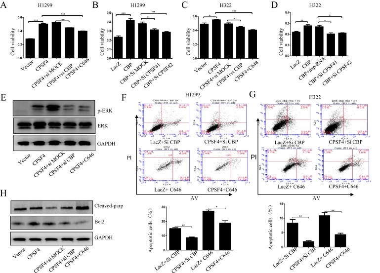
Figure 6.

The synergistic regulation of lung cancer cell growth and apoptosis by CBP and CPSF4. (A, C) Cell viability analysis in H1299 cells stably expressing CPSF4 after transfection with CBP siRNA or treatment with C646. (B, D) Cell viability analysis in H322 cells after co‐transfection with CBP‐overexpressing plasmids and CPSF4 siRNAs. (E) Western blot analysis of the ErK and p‐ErK expression in H1299 cells stably expressing CPSF4 after transfection with CBP siRNA or treatment with C646. (F–G) Apoptosis assay in H1299 and H322 cells after co‐treatment respectively with Lac Z plasmids and CBP siRNA, or Lac Z plasmids and C646, or CPSF4 plasmids and CBP siRNA, or CPSF4 plasmids and C646. The corresponding quantitative analysis of the apoptotic cell numbers was given below. (H) Western blot analysis of the Bcl‐2 and cleaved PARP expression in H1299 cells stably expressing CPSF4 after transfection with CBP siRNA or treatment with C646. Data In panel (A–D) are all represented as mean ± SD of three separate experiments with statistic significance calculated from the two‐tailed student's t test. (*P < 0.05, **P < 0.01).
