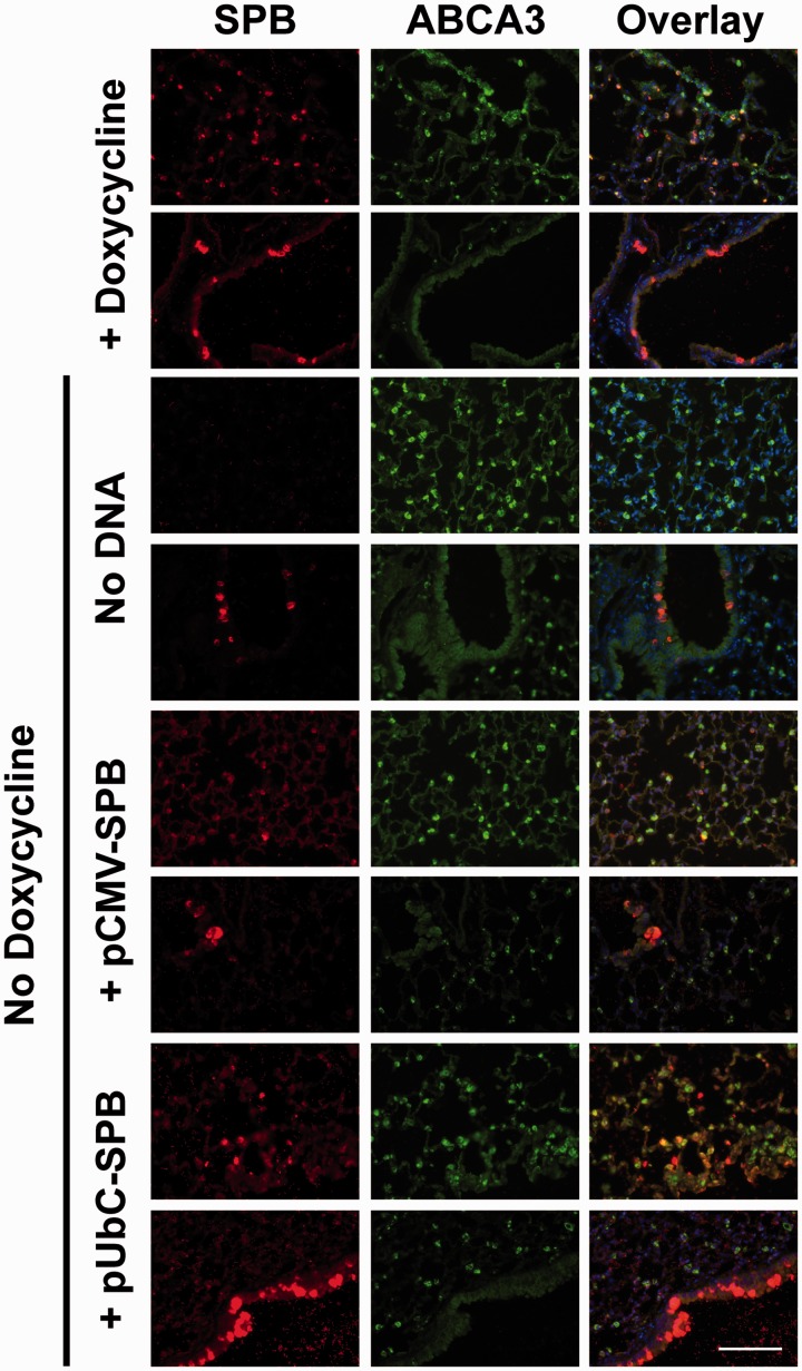
Figure 2.
Immunofluorescent staining of SP-B in compound SP-B mice maintained on doxycycline or following gene transfer and removal of doxycycline. Compound SP-B transgenic mice (n = 4) were either maintained on doxycycline or were taken off doxycycline and 100 µg of each plasmid was delivered to the lungs by electroporation as in Figure 1. Four days later, lungs were removed from animals and inflation-fixed for paraffin embedding and thin sectioning. Sections were reacted with antibodies against SP-B (red) and ABCA3 (green), a marker of alveolar epithelial type II cells, followed by fluorescently-labeled secondary antibodies. Sections were reacted with DAPI (blue) to visualize nuclei. Two representative fields are shown for each experimental group. All images were taken at the same exposure and settings for each antibody. Bar = 100 µm

