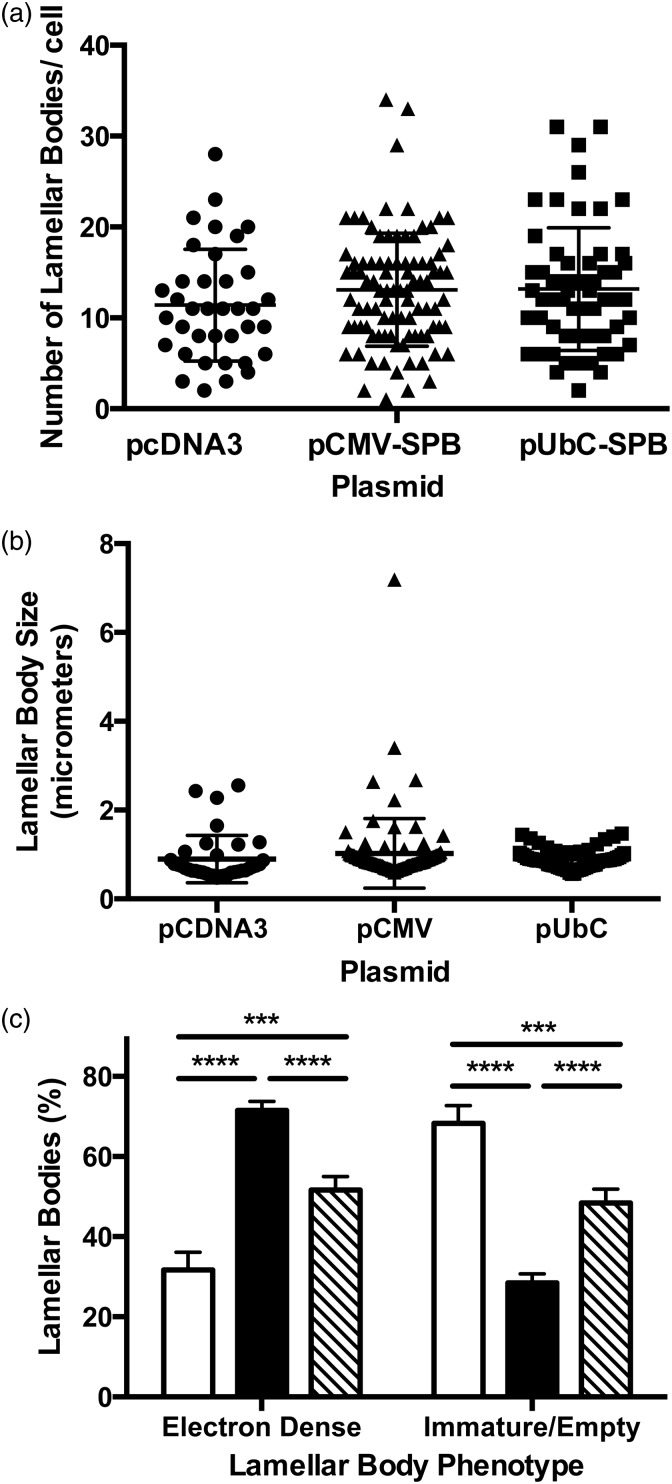
Figure 6.
Analysis of lamellar body phenotypes in mice following gene transfer. Compound SP-B transgenic taken off doxycycline and electroporated with pcDNA3, pCMV-SPB, or pUbC-SPB as in Figure 5 were analyzed for lamellar body phenotype. Between 70 and 110 type II pneumocytes were imaged at 15,000× magnification for each condition. The number of lamellar bodies in each cell was counted and their phenotype assessed as either containing packed electron-dense, concentric surfactant (“electron-dense”), or having a bubbly appearance with no organized lamellae as previously described (“immature/empty”).18 The total number of lamellar bodies in each cell (a), the average size of lamellar bodies in each cell (b) and the phenotype of lamellar bodies in each condition (c) were measured. Open bars represent mice receiving pcDNA3, closed bars represent those receiving pCMV-SPB, and hatched bars represent those receiving pUbC-SPB. ***P < 0.001 and ****P < 0.0001 by 2-way ANOVA and post hoc Tukey multiple comparisons test

