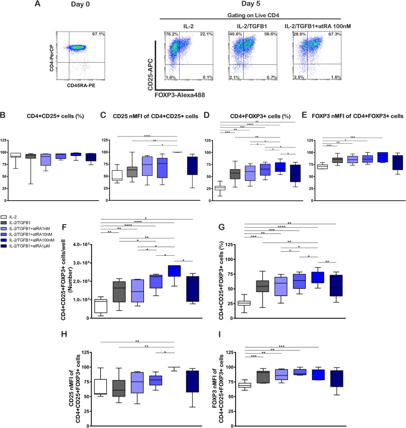
Fig 1. atRA enhances the generation of TGF-β1/IL2-induced Treg cells from naive T cells.
Purified naive T cells (CD4+CD45RA+) were stimulated on day 0 with anti-CD3/CD28 coated beads in the presence of IL-2 (100U/mL), TGF-β1 (5ng/mL), and different concentrations of atRA. Cell phenotype was analyzed by flow cytometry on day 5. (A) Representative dot plots of one donor show the expression of CD45RA+ on the isolated naive CD4 T cells on day 0, and the expression of CD25 and FOXP3 by the generated cells under three different culture conditions on day 5. (B) Percentage of CD25+ cells. (C) CD25 nMFI of CD25+ cells. (D) Percentage of FOXP3+ cells. (E) FOXP3 nMFI of CD25+ cells. (F) Number of CD4+CD25+FOXP3+ Treg cells. (G) Percentage of CD4+CD25+FOXP3+ Treg cells. (H) CD25 nMFI expression of CD4+CD25+FOXP3+ Treg cells. (I) FOXP3 nMFI of CD4+CD25+FOXP3+ Treg cells. Cells were cultured for 5 days in presence of IL-2 (100U/mL), TGF-β1 (5ng/mL) and different concentrations of atRA as shown. Statistical significance was determined by two-tailed paired t tests. *p < 0.05; **p < 0.01; ***p < 0.001; ****p < 0.0001 for n = 7–8 donors in independent experiments. Gating strategy and representative dot plots are shown in S1 Fig.

