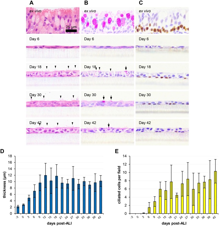
Fig 1. Histological assessment of ovine tracheal epithelial cell culture differentiation over time.
Ovine tracheal epithelial cell cultures were grown at an ALI for the indicated number of days (relative to establishment of the ALI), fixed and paraffin embedded using standard histological techniques. Samples of ex vivo tissue were dissected from the center of each trachea prior to cell extraction, fixed, embedded and processed. Sections were taken, deparaffinized and stained as follows. (A) H&E staining of tissue layers at the indicated time points; selected ciliated cells are indicated by arrowheads. (B) PAS staining to detect mucus-containing/secreting cells (indicated by arrows and arrowheads). (C) Labelling of the transcription factor p63 to detect basal stem cells (positively labelled cells possess brown labelled nuclei). (D) Cell layer thickness was measured using ImageJ. Five images (400× magnification) were taken per insert with three points being measured per image. (E) The numbers of ciliated cells per field were counted from five images per insert. Three inserts were analysed per time point and the data represented is the mean +/- standard deviation from tissues derived from three independent animals (D and E). One-way ANOVA with post-test for linear trend was performed on data with significant (P<0.001) increasing trends being observed for both thickness (D) and ciliation (E).

