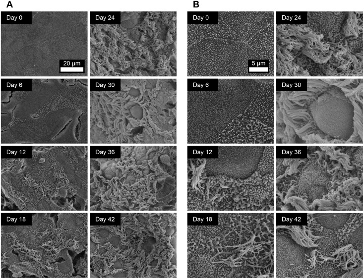
Fig 3. Ultrastructural analysis of the apical surface of ovine tracheal epithelial cell cultures by SEM.
Ovine tracheal epithelial cell cultures were grown at an ALI for the indicated number of days (relative to establishment of the ALI), fixed and processed for SEM. (A) Images were taken at 1500× magnification. (B) Images were taken at 5000× magnification. Ciliated epithelial cells were observed from day 12 onwards.

