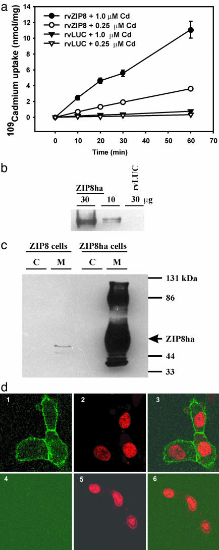
Fig. 3.
Increased Cd uptake caused by membrane-localized ZIP8. (a) Time/dose-dependent 109CdCl2 uptake into rvZIP8 or rvLUC cells. (b) Western blot of ZIP8ha in microsomes (30 vs. 10 μg per lane). (c) Western blot of ZIP8ha in cytosol (C) or microsomes (M) (30 μg per lane) from rvLUC or rvZIP8ha cells. Arrow denotes band at 55 kDa. (d) Localization of the ZIP8ha protein. rvZIP8ha cells (Upper) or rvLUC cells (Lower) were fixed and incubated with a primary anti-HA antibody and a secondary goat anti-rabbit FITC-conjugated antibody. Cells were counterstained with propidium iodide (PI) to visualize nuclei. Confocal fluorescent microscopy detected FITC (Left), PI (Center), or both FITC and PI (Right).

