
Figure 7.
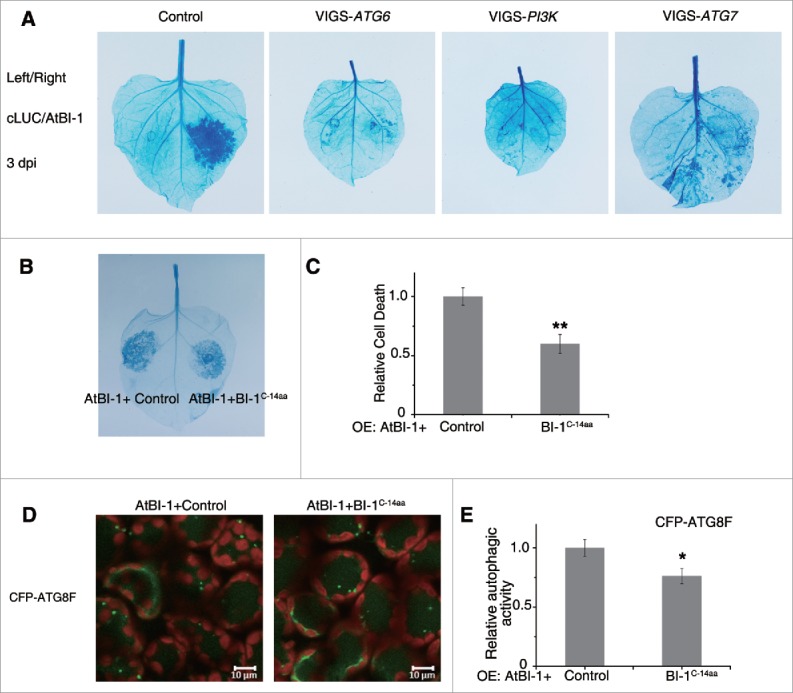
The prodeath role of plant BI-1 required the autophagy pathway. (A) Silencing of ATG6, PI3K or ATG7 suppressed the cell death induced by overexpression of AtBI-1. 2 × 35S-Ω:AtBI-1 was transiently expressed in the silenced leaves (VIGS-ATG6, VIGS-PI3K and VIGS-ATG7) or the nonsilenced control leaves (Control). At 3 dpi, leaves were detached for trypan blue staining. The experiment was repeated at least 3 times, using 5 or more plants per experiment. (B) Overexpression of C-terminal 14 aa of BI-1 decreases AtBI-1 triggered cell death. 2 × 35S-Ω:AtBI-1 was coexpressed with 35S:cLUC-3xHA (Control) or 35S:cLUC-NbBI-1C-14aa-HA (BI-1C-14aa) by agroinfiltration. Trypan blue staining was performed after 48 hpi. (C) Quantitative representation of AtBI-1-induced cell death. To quantify death intensity, images were converted to gray scale, and mean gray value of inoculation area was scored after subtracting the background of noninoculation area by ImageJ. Data represent means ± SEM (**P < 0.01, n = 6). The experiments were repeated 2 times. (D) Representative images of CFP-ATG8F-labeled autophagic structures from leaf tissues agroinfiltrated with 2 × 35S-Ω:AtBI-1, 35S:cLUC-3xHA (Control) or 35S:cLUC-NbBI-1C-14aa-HA (BI-1C-14aa) at 42 hpi. The puncta in the mesophyll cells were autophagic structures. (E) The relative autophagic activity in CFP-ATG8F-labeled tissue. The number of CFP-ATG8F-labeled autophagic structures per leaf section was counted and normalized to control. Data are the means±SEM of relative autophagic activity, with the mean value of the control set to 1.0 (*P < 0.05, n = 30). Experiments were repeated at least 2 times. All autophagy-related experiments were performed with E64d treatment.