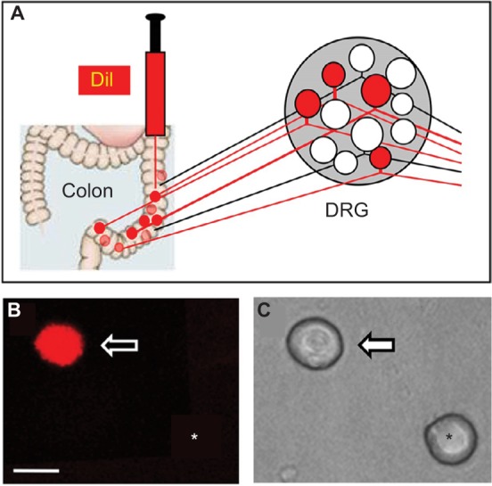
Figure 2.

Retrograde labeling of DRG neurons innervating the colon.
Notes: (A) Fluorescent dye DiI was injected into the colon wall, which helped trace, in a retrograde manner, DRG neurons innervating the colon. (B) Example of a Dil-labeled DRG neuron (arrow). Asterisk indicates the place where a neuron is not labeled by DiI. (C) Phase image of the same DRG neuron labeled by DiI is shown on the left (arrow) and the neuron not labeled by DiI is shown on the right (*). Scale bar: 25.0 μm.
Abbreviation: DRG, dorsal root ganglion.
