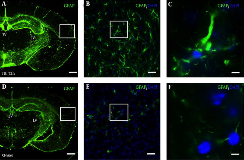
Figure 4. Robust Astrocyte Reactivity Observed in the Ipsilateral S1BF Cortex After Blast.

Brain slice from a TBI mouse adjacent to the TRD slices from Figure 3A shows robust GFAP staining in the A, S1BF region compared to an adjacent brain slice from Figure 3D from D, a SHAM mouse; A and D, both imaged at 2.5×, scale bar = 200 μm; White box in panel A outlines panel B showing astrocyte reactivity with GFAP at 20×, B, scale bar = 30 μm; white box in panel B outlines panel C showing robust astrocytic processes at 63×, C, scale bar =10 μm; White box in panel D outlines panel E showing weak GFAP staining at 20x, E, scale bar = 30 μm; white box in panel E outlines panel F showing weak GFAP staining at 63×, F, scale bar =10 μm. GFAP = glial fibrillary acidic protein; DAPI = 4′,6-diamidino-2-phenylindole (nuclear stain).
