
Figure 1.
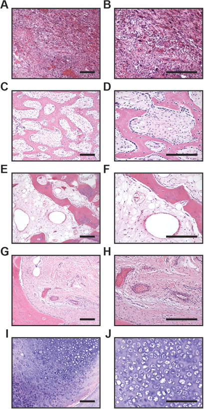
Representative histologic images of heterotopic ossification. (A,B) Typical histologic images of areas with nodular fasciitis-like appearance. Lesion is highly cellular and composed of sheets and fascicles of spindled cells, with scattered multinucleated giant cells, rare foci of bone matrix formation, and numerous slender, elongated capillary-type vessels. (C,D) Typical areas of HO with woven bone. Decalcified section demonstrates anastomosing trabeculae of woven bone with prominent osteoblast lining. The intervening fibrous stroma houses thin-walled capillaries and veins. (E,F) Typical areas of HO with lamellar bone. Decalcified sections composed of thickened trabeculae of lamellar bone. The intervening stroma is composed of fibrous and adipose tissue, with increasing numbers of dilated vessels resembling sinusoids of the bone marrow. (G,H) Typical appearance of outer aspect of HO with fibrous capsule immediately peripheral to lesional tissue. Dense collagenous tissue houses a mixture of thick-walled arteries as well as veins. (I,J) Typical foci of cartilage and endochondral ossification, with rare vessels predominantly at the periphery of cartilage lobules. Scale bar: 100 um.