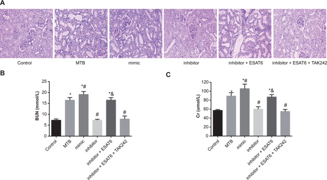
Figure 1. Comparisons of pathological changes of renal tissues of mice amongst groups.
(A) HE (Hematoxylin and Eosin) staining of renal tissues amongst groups (×400); (B) detection of BUN; (C) detection of Scr; *, compared with the control group, P<0.05; #, compared with the MTB group, P<0.05; &, compared with the inhibitor group, P<0.05.

