Fig. 5.
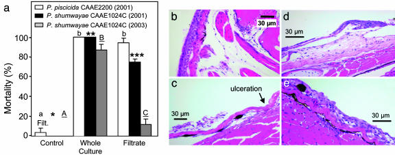
Experiments with fish in whole cultures of Pfiesteria versus in culture filtrate. (a) Juvenile tilapia killed (percent) by whole cultures and filtrate of P. shumwayae CAAE1024C and P. piscicida CAAE2200 from positive SFBs (means ± 1 SE; n = 10 fish per treatment; repeated 6 time for whole cultures, 10 times for filtrates; letters and asterisks indicate significant differences (P < 0.01 to 0.05]; modified from ref. 11). P. piscicida and P. shumwayae 2000–2002 tests were compared with retests in 2003. The P. piscicida strain had lost toxicity (no fish death in whole-culture SFBs or filtrate; data not shown). The P. shumwayae strain had lower toxicity [caused some death in whole-culture SFBs and filtrate (24)]. (b) Skin and underlying musculature from an unexposed control fish (pectoral area, epithelial cells four to six cell layers thick). At the dermal–epidermal interface there was mild artifactual separation, accentuating the stratum spongiosum. Deep to the skin were normal subcutis, skeletal muscle bundles, and bone. (c–e) Eroded and ulcerated skin from fish exposed for 8 h to cell-free filtrate from toxic P. shumwayae CAAE1024C, showing necrosis and fragmentation of eroded epidermis (lifted from the basement membrane) (c); dermal–epidermal cleft (blister-like) with underlying area of severe dermal edema that expanded the stratum spongiosum (d and e). Necrosis of Malpighian epithelial cells in the epidermis and dermal edema suggest that these changes were not artifacts of handling, immersion fixation, or processing. (e) After 4 h, increased edema between Malpighian epithelial cells and spongiosis. Occasional apoptotic cells and intracellular edema are consistent with early epithelial injury.

