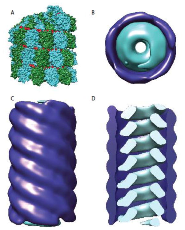
Figure 2.
Organization of the paramyxovirus nucleocapsid. A) Nucleocapsid proteins for all paramyxoviruses form helical assemblies of N proteins encapsidating the viral RNA (shown in this model are MeV RNPs, N proteins are depicted in cyan and forest green, the RNA is colored in red) (PDB ID: 4UFT). B–D) While RNPs for 20 nm diameter tubules, MeV RNPs were also found M protein wrapped in larger diameter 30 nm tubules [43]. Matrix proteins are shown in dark blue and nucleocapsids in cyan. Top view (B) and side view (C) of the 30 nm tubules depicting the distinct cylindrical M complex surrounding the MeV nucleocapsid. D) A clipped model of the 30 nm tubule structure. The matrix coated nucleocapsids were created using Chimera [92] based on electron density maps EMD-1973 and EMD-1974 [43].

