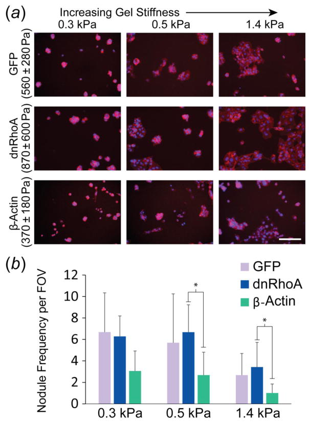
Figure 3.
Effect of mechanophenotype on cellular assembly into nodules or plaques. (a) Representative images of transfected WI-38 cells on PAAm gels (nuclei: blue, actin filaments: red; scale bar: 200 μm). (b) Abundant nodule formation occurred on gels that were more compliant than the inherent mechanophenotype of cells in the noted cell line. Nodule frequency per field of view (FOV) shown as mean ± s.d. (* p < 0.05, as determined by a two-factor ANOVA between cell type and gel stiffness on logarithmically transformed data, followed by Holm-Sidak post-hoc analysis).

