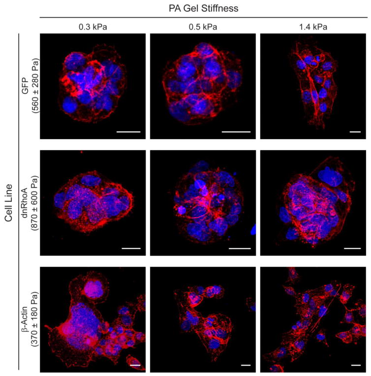
Figure 5.
Nodules and plaques of GFP, dnRhoA, and β-Actin cells after 4 days of culture. Confocal projections revealed differences in actin bundle formation (nuclei: blue, actin filaments: red; scale bars: 20 μm) on gels, especially as cells transitioned from nodule to plaque formations on PAAm gels greater than the mechanophenotype of the adhered cells.

