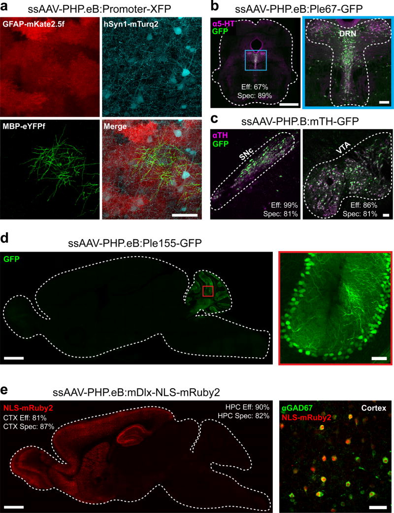
Figure 5. AAV-PHP.eB can be used with gene regulatory elements to achieve cell type-restricted gene expression throughout the brain.
(a) Images show co-transduction of the cortex 3 weeks after co-administration of 3 vectors with 3 different promoters driving distinct XFPs (intravenous injection of AAV-PHP.eB at 1 × 10¹² vg per viral vector). (b) A vector providing GFP expression driven by a FEV/serotoninergic neuron-specific promoter (ssAAV-PHP.eB:Ple67-GFP) was intravenously delivered at 1 × 10¹² vg and co-localized to serotonin (5-hydroxytryptamine, 5-HT, magenta) expressing cells in the dorsal raphe nucleus (DRN) outlined in blue and expanded for detail (right). (c) A vector providing GFP expression from a mouse tyrosine hydroxylase (TH) promoter (ssAAV-PHP.B:mTH-GFP) was intravenously injected at 1 × 10¹² vg, and imaging with IHC for TH (magenta) was performed after 2 weeks of expression. Images show the substantia nigra pars compacta (SNc, left) and ventral tegmental area (VTA, right). (d) A vector with a Purkinje cell-selective promoter (Ple155, Pcp2) driving GFP (ssAAV-PHP.eB:Ple155-GFP) was intravenously injected at 1 × 10¹² vg and expression was examined at 4 weeks. A whole sagittal section (left) shows native GFP fluorescence (green) in the cerebellum (left) in cells with the morphology of Purkinje cells (see higher resolution of the red boxed region, right). (e) A vector with a forebrain GABAergic interneuron-specific enhancer driving nuclear-localized mRuby2 (AAV-PHP.eB:mDlx-NLS-mRuby2) was intravenously injected at 3 × 10¹¹ vg and expression was examined at 8 weeks (CTX: cortex, HPC: hippocampus). Native mRuby2 expression within the forebrain (red, left). Co-localization was assessed by IHC for GABAergic cells (GAD67+, green, right). The scale bars in (a; b, right; c; d, right; e, right) are 50 µm. For (b and d, left) the scale bars are 1 mm. All panels are confocal images of native XFP fluorescence. The efficiency (Eff) and specificity (Spec) values for transduction by FEV/Ple67, mTH, mDlx vectors are giving in the DRN, VTA, SNc, cortex (CTX), and hippocampus (HPC).

