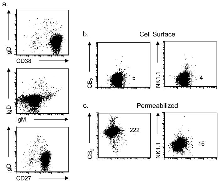
Fig. 3. A malignant B cell line expressing an activated phenotype exhibits the same pattern of CB2 expression as that observed with primary activated B cells from tonsils.
(a) Cells from the malignant B cell lymphoma cell line, SUDHL-4, exhibited cell surface markers consistent with an activated B cell subset based on the expression pattern for IgD (negative), CD38 (positive), and CD27 (positive). (b) Cells were stained while still viable for detection of cell surface CB2 with a primary unlabeled mAb against CB2 protein or isotype control, NK1.1, and then stained with APC-labeled GAM. (c) For total cell expression of CB2, cells were fixed and permeabilized prior to specific staining. Numbers represent relative MFI for staining on the Y axis. Representative experiment shown, n = 3.

