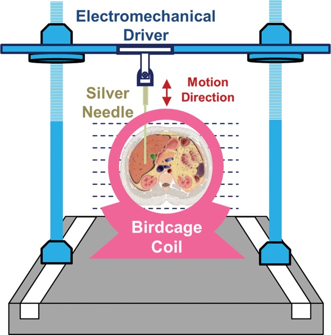
Figure 1a:
(a, b) Experimental set-up of liver MR elastography imaging in mice (a) and pigs (b) respectively. (c) In vivo multifrequency three-dimensional wave images and elastograms. Wave images at multiple frequencies indicate good shear wave propagation throughout the majority of the liver. The three orthogonal motion directions refer to x (frequency-encoding direction), y (phase-encoding direction), and z (section direction). Elastograms were obtained with the three-dimensional direct inversion algorithm. Transverse magnetization (anatomic) images of a mouse with NAFLD (coronal plane) and a fumarylacetoacetate hydrolase pig (axial plane) are shown in the right column. The white dot in the center of the mouse liver is where the silver needle was applied. The color maps used for the wave and stiffness images are also shown in this column.

