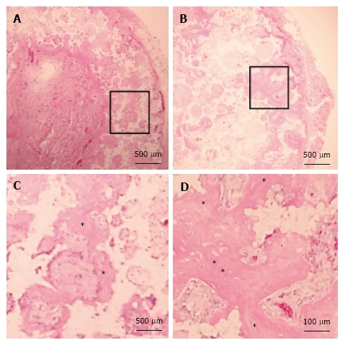
Figure 6.

Histology of β-tricalcium phosphate discs wrapped with osteogenic matrix cell sheets. Cell sheets were exposed to hypoxia (5% O2) for 7 d followed by normoxia (21% O2) for 7 d (B) or normoxia for 14 d (A). Prominent newly formed bone (*) was observed in the HN group. (C) and (D) are higher magnifications of squared areas indicated in (A) and (B), respectively.
