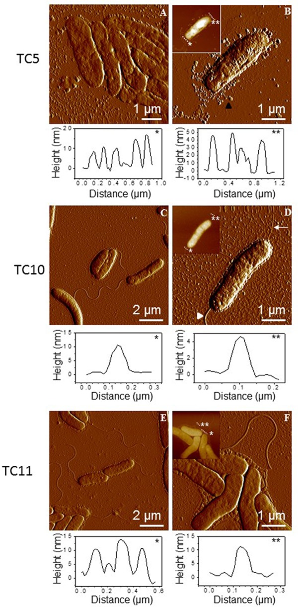
FIGURE 4.

Imaging bacterial surface in air. AFM images of TC5 (A,B), TC10 (C,D), and TC11 (E,F) were performed in air using contact mode. AFM deflection and height (insets) images of post-exponentially growing cells that were directly deposited on mica and dried prior analysis. Vertical cross sections taken in the height images (asterisks indicate the correspondence with dashed lines) are also shown to emphasize sizes of cellular structures.
