
Figure 6.
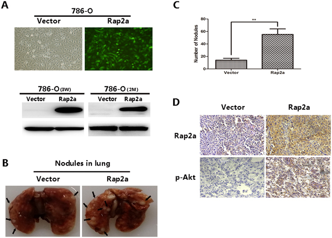
Rap2a enhances RCC cell metastasis in vivo. (A) 786-O cells were examined for their infection efficiency under the fluorescent microscope after lentivirus transfection (×100 magnification) (top panel). Western blot of Rap2a from Rap2a-overexpression 786-O cell lines and Ctrl-786-O cell lines selected with puromycin for 3 weeks after lentivirus infection. Rap2a expression levels remain stable without puromycin selection for 2 months (bottom panel). (B) Representative images of 10% buffered formalin fixed lungs with metastatic nodules 2 months after respective injection of Ctrl, Rap2a-overexpression 786-O cell lines. Arrows indicate metastatic nodules. The number of lung metastatic nodules was counted under a dissecting microscope. (C) A statistically dramatic increase in the number of the lung metastases was seen in Rap2a-overexpression group compared with the Ctrl group. Data are displayed with means ± SD from 10 mice in each group. (D) Immunostaining of Rap2a and p-Akt in metastatic nodules of Rap2a-overexpression and Ctrl 786-O groups. Rap2a and p-Akt expression in Rap2a-overexpression group were much higher compared with Ctrl group. **P < 0.01.