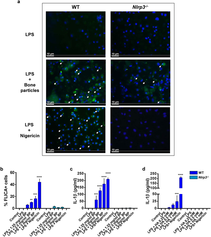
Figure 2.

Bone matrix components provide secondary signals that assemble functional inflammasomes. WT or Nlrp3 −/− BMM were incubated for 3 hours with 100 ng/ml LPS and stimulated with 15 µM nigericin for 30 minutes, bone particles (BP, 1.25–5 mg/ml) or hydroxyapatite (HA) crystals (0.25–1 mM) for 1 hour. (a) Cells were then incubated with FLICATM FAM-YVAD-FMK probe and analyzed by fluorescence microscopy. Original magnification, 20x; scale bars = 10 µm; white arrows show foci of caspase-1 activation. (b) Quantification of FLICA+ cells. Data are means ± SD of at least 3 fields. (c,d) IL-1β release in the conditioned media. Data are means ± SD from experimental triplicates, and are representative of at least 2 independent experiments. (b) **p = 0.0062, ***p = 0.0003, ****p < 0.0001; (c) ***p = 0.0008, ****p < 0.0001. p values correspond to significant differences relative to the control for each genotype.
