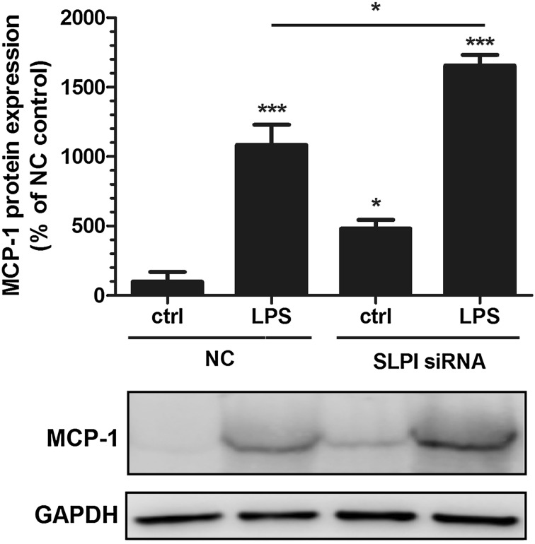
Fig. 5.
Knockdown of SLPI by siRNA enhances PDL cell expression of MCP-1 protein. Both basal and LPS-stimulated levels of MCP-1 protein are elevated in PDL cells treated with SLPI siRNA compared to NC-treated control cells. The cells were transfected with either SLPI siRNA or NC construct for 96 h and treated with or without 1 µg/ml LPS during the last 24 h of the 96 h transfection period. The MCP-1 protein was determined in PDL cell lysates by Western blotting. MCP-1 is detected as one band at the expected molecular weight (12–13 kDa). The relative MCP-1 concentration was assessed by densitometry and normalized to the GAPDH serving as internal control. Values are presented as mean ± SEM of 4 observations in each group. * and *** represent P < 0.05 and P < 0.001, respectively, versus NC ctrl. * represents P < 0.05 for the comparison indicated by the horizontal line

