
Figure 5.
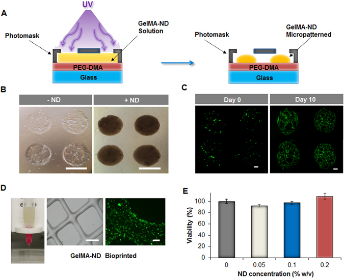
Microfabricated structures using GelMA/ND nanocomposite hydrogels and evaluation of their biocompatibility. (A) Schematic describing the process of micropatterning of the GelMA/ND prepolymer solution into nanocomposite hydrogel with a 1 mm diameter. (B) Brightfield images of 1 mm in diameter micropatterned hydrogels with (+ND) and without (−ND) NDs encapsulating hASCs in 3D after ten days of culture. (Scale bar = 1 mm). (C) Calcein-AM staining of hASCs cultured in GelMA/ND micropatterned hydrogels at different time points. hASCs proliferated and spread throughout the microfabricated constructs after ten days. (Each scale bar = 200 µm). (D) The prepolymer GelMA/ND solution can also be bioprinted into more complex structures as shown in the phase contrast image (scale bar = 1 mm). The calcein-AM staining (green) shows the hASCs that were seeded on the bioprinted nanocomposite GelMA/ND and cultured for five days. (Scale bar = 100 µm). (E) MTS assay of hASCs encapsulated in GelMA/ND nanocomposite hydrogels demonstrated excellent biocompatibility regardless of the ND concentration in the gel. Results are reported as mean ± S.D., (n = 5). No statistical difference was observed among the groups tested.