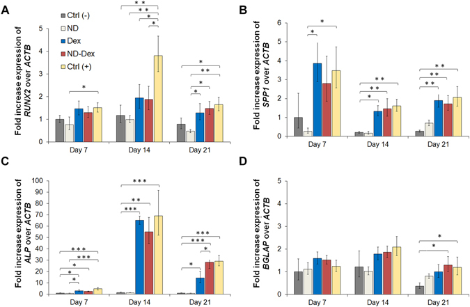
Figure 7.

Assessment of osteogenic differentiation of hASCs by qPCR analysis. The fold expression increase of different genes have been evaluated including (A) RUNX2, (B) osteopontin SPP1, (C) alkaline phosphatase ALP and (D) osteocalcin BGLAP. β-actin ACTB has been used as control gene and data were calculated following the relative method ΔΔCt. Results are normalized based on the values obtained from the Ctrl (−) group at day 7. Groups tested are respectively hASCs cultured using GelMA hydrogels in basal media Ctrl (−), GelMA/ND 0.2% w/v nanocomposite scaffold without Dex in basal media (ND), GelMA hydrogels containing dexamethasone 1 µM in osteoconductive media (Dex), the nanocomposite system containing the complex in osteoconductive media (ND/Dex) and hASCs cultured in osteoinductive media Ctrl (+). Results are shown as mean ± S.D. (n = 3) (*p < 0.05 **p < 0.01, ***p < 0.001).
