Figure 4.
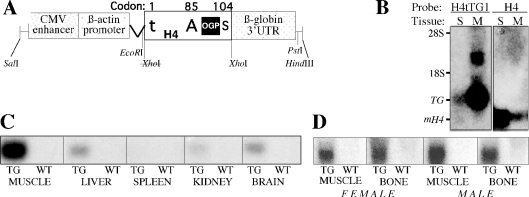
(A) Schematic illustration of the H4tTG1 transgene. The transgene is composed of the human CMV enhancer, the chicken β-actin promoter and intron, a rat histone H4 coding sequence and the rabbit β-globin 3′-UTR. Within the H4 box, t represents the mutation in the H4 translational initiator (from ATG to tTG at codon #1), A represents the alternative ATG initiation codon (#85), giving rise to preOGP, S represents the stop codon (#104) and the black square depicts the OGP-coding sequence (H490–103). (B) H4tTG1 transgene versus endogenous H4 expression. An aliquot of 20 μg RNA from the spleen (S) and muscle (M) of a 17-week-old transgenic mouse were subjected to northern analysis using as probe either the rabbit β-globin 3′-UTR or a murine histone H4 sequence. 28S and 18S, location of the ribosomal RNA bands; TG, transgenic mRNA; and mH4, endogenous murine H4 mRNA. (C and D) Ubiquitous H4tTG1 transgene expression. Transgene expression was measured by northern analysis of 20 μg RNA from the indicated tissues of a transgenic (TG) and a wild-type (WT) mouse using the rabbit β-globin 3′-UTR as probe. In (D), the RNA was obtained from either a female or a male as indicated.

