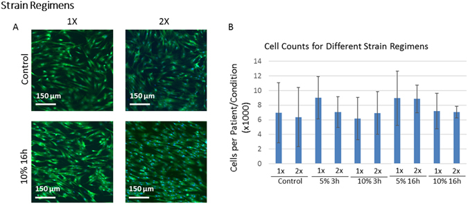
Figure 4.

Shape and Cell Count of Stretched and Unstretched MSCs. Stretched and unstretched MSCs that adhered to the collagen type I sheets were stained with Calcein to illustrate MSC shape (A). Cell nuclei stained with DAPI (not shown) were counted to measure cell numbers (B). The axis of stretch was along the horizontal direction. Clear differences in MSC alignment can be seen in stretched vs. unstretched MSCs, and alignment was more pronounced in 2× than 1× stretched MSCs (A). More subtle differences in cell size and cell spreading were also present and quantified elsewhere. The number of MSCs across the experimental conditions tested was not statistically different. Error bars indicate standard deviation.
