
Figure 4.
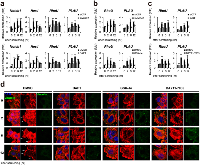
Notch1 regulates keratinocytes migration via RhoU and PLAU gene expression in scratch-wounded keratinocytes. (a) Depletion (siNotch1) or inactivation (DAPT) of Notch1 (b) Depletion (siJMJD3) or inactivation (GSK-J4) of JMJD3 (c) Depletion (sip65) or inactivation (BAY 11-7082) of NF-κB suppresses RhoU and PLAU gene activation in scratch-wounded HaCaT keratinocytes. Transcripts of Notch1, Hes1, RhoU, PLAU, and GAPDH were determined by quantitative PCR. (d) Inactivation of Notch1 or JMJD3 or NF-κB results in decreased filopodia-like protrusions (white arrows), increased focal adhesion and actin stress fibers in scratch-wounded HaCaT keratinocytes. To detect filopodia and actin stress fibers, cells were stained with phalloidin (red). Focal adhesion was identified by cell immunostaining with anti-vinculin antibody (green). Nuclei were identified using DAPI staining. Scale bar, 25 μm.