Figure 2.
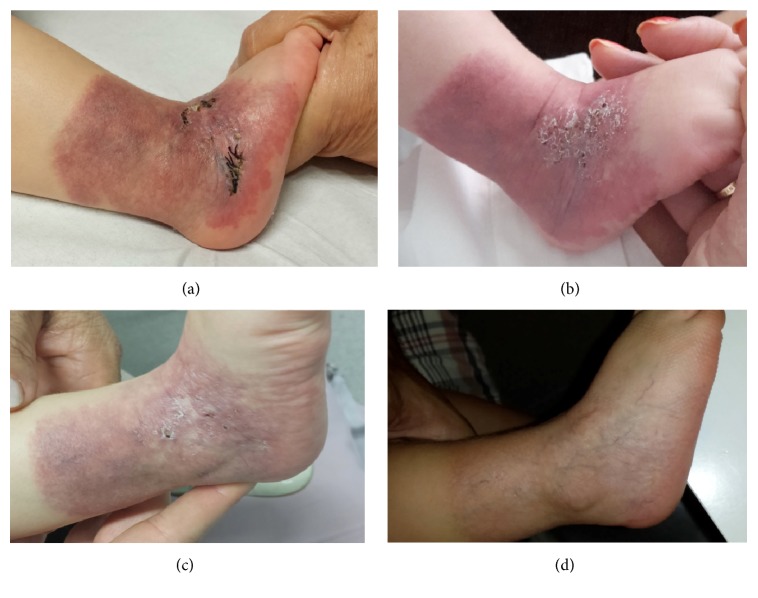
Case 2. (a) A skin biopsy on erythematous scaly plaque with geometric border covering all lateral aspect of her right ankle as a sock-like distribution. (b) Numerous superficial ulcerations of this erythematous scaly plaque. (c) Progressive regression of the lesion and the ulceration with propranolol treatment. (d) Spontaneous involution of the lesion.

