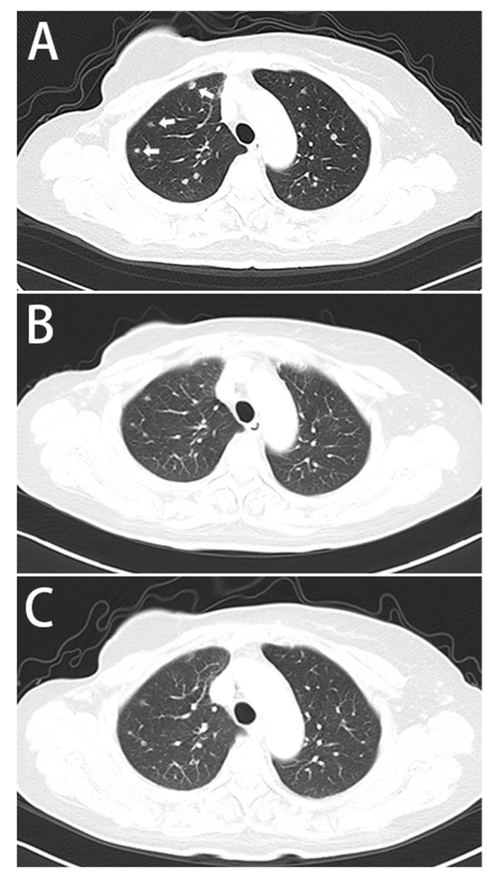
Figure 2.

CT scan demonstrating the progression of the pulmonary metastases. (A) CT scan of the chest (December 2013) revealing increased number of nodules in the lungs, indicated by arrows. (B) CT scan (May 2014) revealing partial remission of the lesions in the lungs. (C) CT scan (December 2015) demonstrating stable disease. CT, computed tomography.
