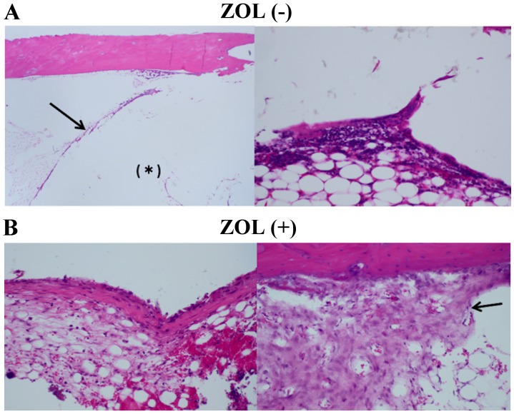
Figure 8.
Histopathology of bone tissue surrounding implanted bone cement. (A) Control bone cement was packed into the tibia bones of rabbits. *Internal side of the arrowhead indicates the area filled with bone cement (B) ZOL-loaded bone cement was packed into tibia bones of rabbits. Bone cement was covered with fibrous tissue, which is lined with multinucleated giant cells and lymphocytes. Fibrous tissue was slightly increased with ZOL-treatment. Proliferated osteoid tissue is observed at a distance from the bone cement (black arrow). There was no obvious osteonecrosis observed compared with control bone cement treatment. ZOL, zoledronic acid.

