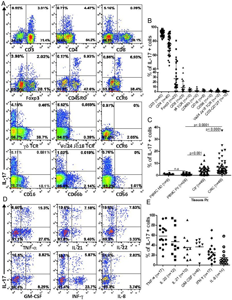
Figure 2.
Colorectal cancers (CRC)-infiltrating interleukin (IL)-17+ cells are polyfunctional Th17. Single cell suspensions from freshly excised clinical specimens of CRC and corresponding tumour-free colonic mucosa (Ctr) and peripheral blood mononuclear cells from healthy donors (PBMC HD) or patients with CRC (PBMC Pz), were incubated with phorbol 12-myristate 13-acetate (PMA)/ionomycin/brefeldin for 5 h. Surface staining for specific cell population markers and intracellular staining for Foxp3 and cytokines was then performed. (A) Representative flow cytometric analysis of CRC infiltrates stained for IL-17 and the indicated cell-specific markers. Tumour-infiltrating cells are gated based on physical parameters, as defined by analysis of autologous PBMC. (B) Frequencies of cells positive for the indicated markers within gated IL-17+ cells. Means are indicated by bars. Numbers of samples evaluated for each marker are indicated in parentheses. (C) Frequencies of IL-17+ cells on gated CD3+ T cells obtained from PBMC HD or PBMC CRC and single cell suspensions from freshly excised clinical specimens of Ctr and corresponding CRC. Means are indicated by bars. Numbers of samples evaluated for each marker are indicated in parentheses. Statistical significance was assessed by t test. (D) Representative flow cytometric analysis of intracellular cytokine staining on gated CRC-infiltrating CD3+CD4+ cells. (E) Frequencies of cells positive for the indicated cytokines gated on CD3+CD4+ IL-17+ T cells. Means are indicated by bars. Numbers of samples evaluated for each cytokine are indicated in parentheses. GM-CSF, granulocyte-macrophage colony stimulating factor; IFN, interferon; TNF, tumour necrosis factor.

