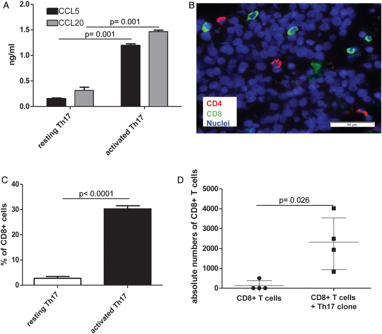
Figure 7.
Th17 cells promote recruitment of cytotoxic CD8+ T cells into colorectal cancer (CRC) tissues. (A) CRC-Th17-infiltrating engineered tumour tissues (see online supplementary figure S10) were left untreated or were activated by adding CytoStim to the perfusion medium. Culture media were collected 20 h later and chemokine contents were assessed by ELISA. Statistical significance was assessed by Mann–Whitney test. (B and C) Three hours after Th17 activation, the perfusion in the bioreactor was stopped and CD8+ T cells were added to the system. After an overnight period, scaffolds were removed and tumour infiltration by CD8+ T cells was evaluated by immunofluorescence analysis upon staining with CD4- and CD8-specific antibodies (B) and by flow cytometry upon staining of single cell suspensions with EpCAM-, CD4- and CD8-specific antibodies (C). Percentages of CD8+ cells in tumour tissues infiltrated by resting or activated Th17 cells are reported. Means±SD from three experimental replicates performed with one Th17 clone are depicted. Statistical significance was assessed by Mann–Whitney test. (D) CSFE+CD8+ T cells were adoptively transferred in tumour bearing mice alone or together with equal numbers of CRC-Th17 (four mice/condition). Absolute numbers of CD8+CSFE+ T cells were evaluated by flow cytometry upon staining of tumour and Th17 cells with anti-EpCAM and anti-CD4 antibodies, respectively. Statistical significance was assessed by Mann–Whitney test.

