Figure 1.

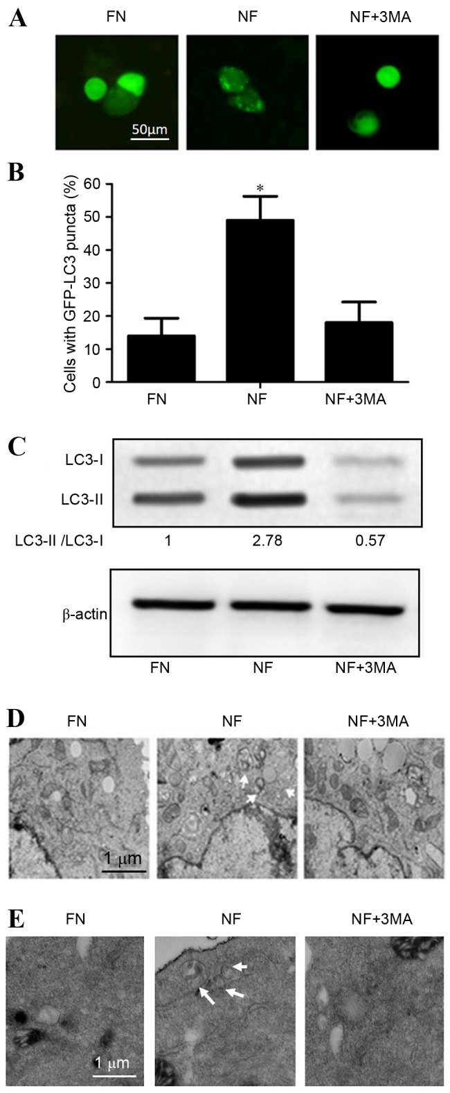
Nutrient-deprived conditions induced autophagy in cholangiocarcinoma cells. Nutrient deprivation conditions induced autophagy in QBC939 and RBE human cholangiocarcinoma cells cultured for 12 h in FN or NF medium, or NF medium containing the autophagy inhibitor 3MA. (A) QBC939 cells were transfected with GFP-LC3 plasmids and GFP-LC3 fluorescence was evaluated by fluorescence microscopy. Representative images are presented. (B) The percentages of GFP-positive cells that exhibited punctate GFP-LC3 fluorescence, indicating the presence of autophagosomes, were evaluated; cells with >2 autophagosomes per cell were scored as exhibiting an autophagic reaction. Data are presented as the mean ± standard deviation of ≥3 independent experiments. *P<0.05 vs. FN group. (C) Whole cell lysates from RBE cells were subjected to western blotting to analyze LC3-I and LC3-II levels. β-actin was included as a loading control. Representative transmission electron microscopy images are presented for (D) QBC939 cells and (E) RBE cells. Arrows indicate autophagosomes. FN, full-nutrient; NF, nutrient-free; 3MA, 3-methyladenine (10 mM); GFP-LC3, green fluorescent protein-tagged LC3; LC3, microtubule-associated protein 1A/1B-light chain 3.
