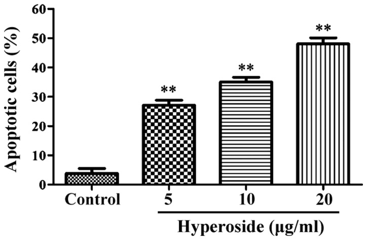
Figure 2.

Percentage of apoptotic cells detected by flow cytometry after after treatment with different concentrations of hyperoside. The apoptosis rates of cells were significantly increased after treatment with hyperoside. **P<0.01 vs. the control group (incubation with 0 µg/ml hyperdoside).
