Figure 4.
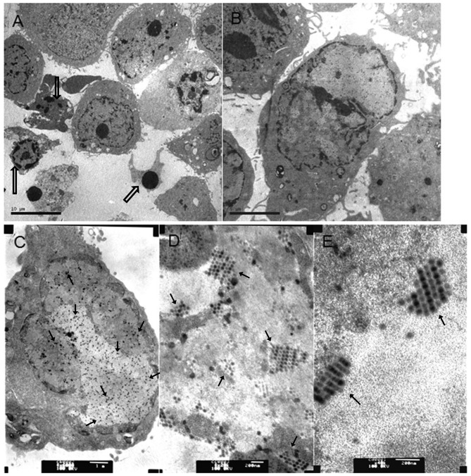
Virus particles in bladder cancer cells infected with Ad5-UPII-E1A or Ad5-UPII-E1 Acombined with MMC or HCPT. (A) A total of 5,637 cells was treated with 0.4 mg/ml HCPT and Ad5-UPII-E1A (10 MOI) for 72 h (magnification, ×3,000). (B) A total of 5,637 cells was treated with 0.2 mg/ml MMC combined with Ad5-UPII-E1A (10 MOI) for 72 h (magnification, ×6,000). (C) A total of 5,637 cells was treated with Ad5-UPII-E1A alone, and the uniform distribution of virus particles within the cell is visible (magnification, ×20,000). (D) Distribution of virus particles within the cell (magnification, ×40,000). (E) Virus particle morphology (magnification, ×40,000). Hollow arrows indicate the fragmented nuclei, the gathering of nuclearchromatin and the formation of apoptotic bodies. Regular arrows indicate the virus particles. Ad5-UPII-E1A, urothelium-specific recombinant adenovirus type 5; MMC, mitomycin; HCPT, hydroxycamptothecin; 10 MOI, 10 multiplicity of infection.

