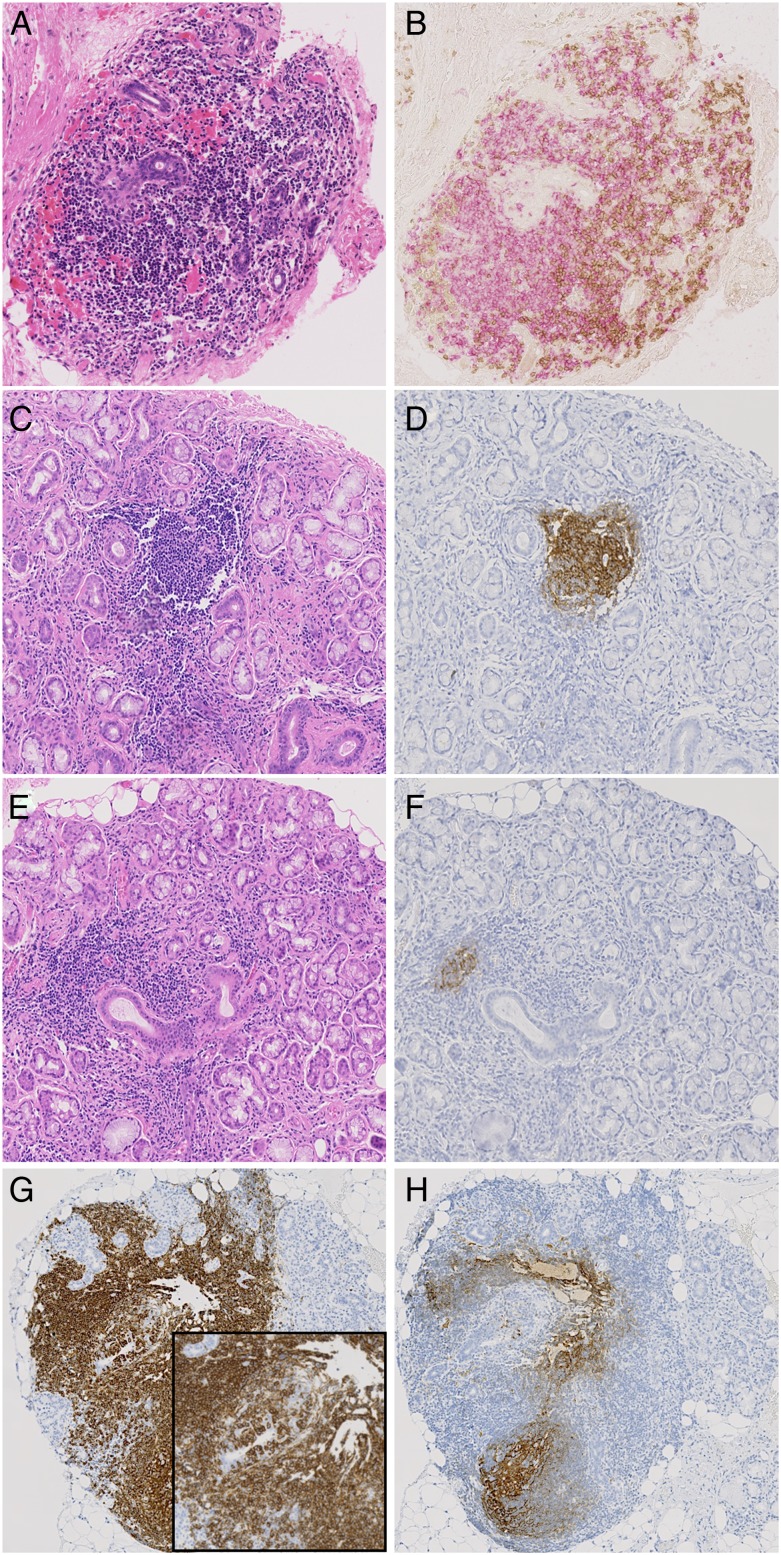
Figure 3.
(A–H) Sequential sections illustrating inflammatory infiltrates in the salivary glands of patients with primary Sjögren's syndrome stained by H&E (A, C, E), CD3 (brown in B), CD20 (pink in B and brown in G) and CD21 (brown in D, F and H). (A and B) Sequential section illustrating segregation in T and B cells in large periductal infiltrate in absence of germinal centre (GC). (C and D) Evident GC in H&E stained section confirmed by CD21 staining on sequential section. (E and F) Small CD21+ cluster of follicular dendritic cells (FDCs) in sequential section of a large aggregate with absence of obvious GC features at the H&E staining. (G and H) Large CD20+ infiltrate with obvious lymphoepithelial lesions (inset) and the presence of CD21+ FDC networks at the sequential section.

