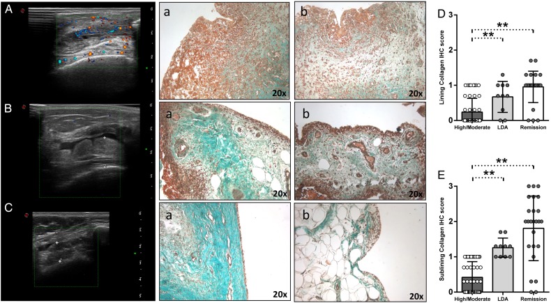
Figure 3.
(A–E) Masson Trichrome Goldner with light green immunostaining on ST of patients with rheumatoid arthritis (RA) in remission, in low disease activity (LDA) and in high/moderate disease. (A) Example photos of collagen (green) staining of ST from high/moderate patient with RA (a, b) (magnification 20×); corresponding ultrasound assessment (US) picture with PD scale (PD score=2) of the knee used for ST biopsy is shown. (B) Example photos of collagen (green) staining of ST from patient with RA in LDA (a, b) (magnification 20×); corresponding US picture with PD scale (PD score=0) of the knee used for ST biopsy is shown. (C) Example photos of collagen (green) staining of ST from patient with RA in remission (a, b) (magnification 20×); corresponding US picture with PD scale (PD score=0) of the knee used for ST biopsy is shown. (D) Lining IHC score for collagen in ST of enrolled cohorts; high/moderate versus LDA patients with RA, p=0.03; LDA versus remission patients with RA, p=0.10; high/moderate versus remission patients with RA, p<0.001. (E) Sublining IHC score for collagen in ST of enrolled cohorts; high/moderate versus LDA patients with RA, **p<0.001; LDA versus remission patients with RA, p=0.10; high/moderate versus remission patients with RA, **p<0.001.

