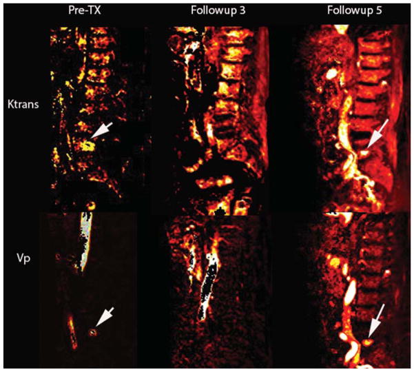
FIG. 5.
Case 1. Sagittal T1-weighted DCE MRI perfusion maps for the parameters Vp and Ktrans before RT, and at follow-up visits 3 and 5 after RT. Follow-up 3 illustrates a hypointensity at L-5, whereas follow-up 5 shows a hyperintensity with Vp values of 5.4 and Ktrans of 1.86. The Vp color map is consistent with the clinical finding of new metastases to L-5. The arrows designate untreated L-4 metastasis prior to radiation therapy.

