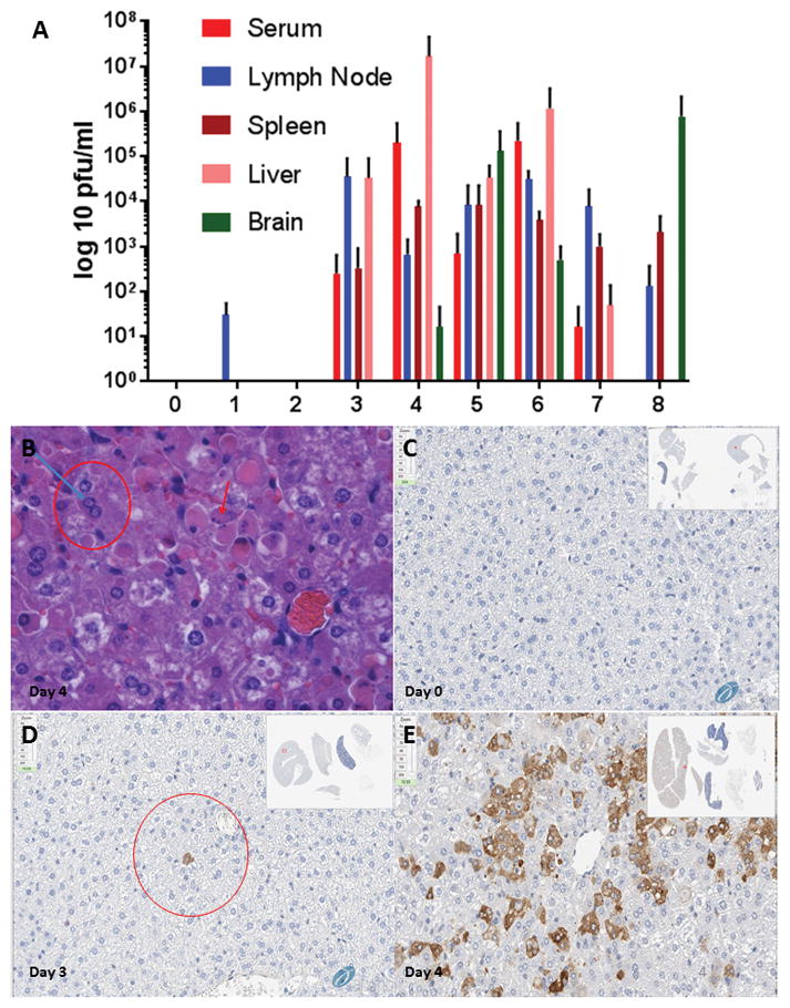
Figure 4. RVFV model validation.

(A) BALB/c mice were randomly assigned to groups of 3 and infected with 1,000 PFU of RVFV ZH501 subcutaneously. Three uninfected control mice were sacrificed on day 0 with infected mice being sacrificed from days 1–8. Liver, spleen, brain, serum, and lymph node were harvested from each mouse and sections were either placed in 10% neutral buffered formalin for histologic evaluation or were used to determine viral titers in each organ via plaque assay. (B) Liver, Infected Mouse, Day 4: Histologic examination revealed groups of hepatocytes with shrunken, hypereosinophilic, cytoplasm and condensed, eccentrically placed nuclei (red arrow) consistent with groups of cells undergoing apoptosis. Adjacent to these areas were nuclei containing 3 to 4 um, hypereosinophilic, intranuclear inclusions (blue arrow/red circle) consistent with RVFV protein inclusions. (C) Uninfected Liver – Day 4, RVFV Immunohistochemistry. (D) Day 3 PI – rare hepatocytes (circle) displayed intracytoplasmic immunohistochemical staining. (E) Day 4 PI – positive staining throughout the liver to include large groups of cells displaying strong, specific, intracytoplasmic immunohistochemical staining.
