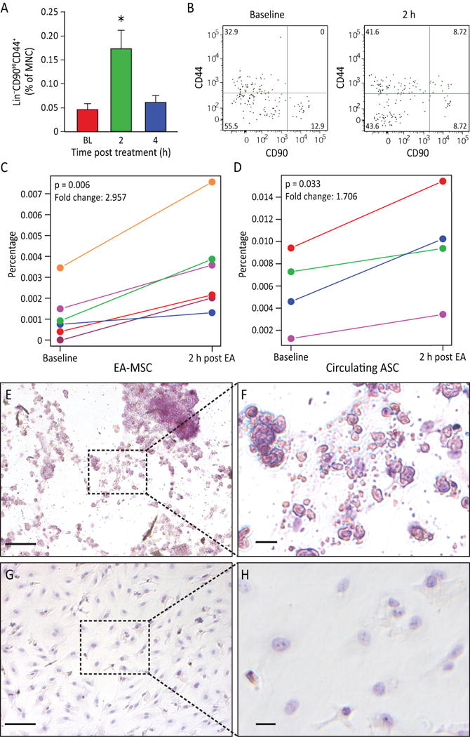
Figure 3. EA stimulation induced MSC stem cell mobilization.

A) Rat peripheral blood MSC were increased (p=0.0063) after EA. Circulating MSC were defined as Lin−(CD45−CD31−erythroid−CD11b−) cells that were positive for CD44 and CD90. Gated cells increased post treatment (n=11 for baseline and 4 hours, n=9 for 2 hours). B) Representative flow charts for rat Lin− cells are shown at baseline and 2 h samples. C) The percentage of human peripheral blood MSC increased in post EA-treatment (p=0.006, n=6). D) The percentage of circulating MSCs from adipose tissue (AD-MSC) are significantly elevated in 2 h post EA-treatment (p=0.033, n=4). E, F) EA-mobilized MSC were expanded in vitro. After undergoing adipogenesis differentiation, EA-mobilized MSC developed fat deposits as seen by Oil Red staining, which were not seen in the undifferentiated control cells (G, H). Magnification bars: E,G: 100 μm; F,H: 50 μm.
