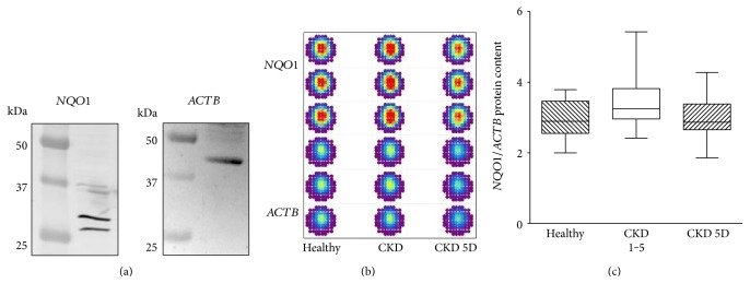
Figure 2.
NQO1 protein content. (a) To show the effective detection of NQO1 and ACTB protein by antibodies in the cell material used in our study, we performed immunoblot analyses with cells obtained from healthy control subjects. Immunoblots of NQO1 (expected size 26/27 kDa and 31 kDa) and ACTB (expected size 42 kDa) in monocytes are shown. (b) Pseudocolored fluorescence intensities of in-cell Western assays for the quantification of the NQO1 protein content relative to the ACTB protein content in monocytes from a healthy subject, a patient with CKD, and a patient with CKD 5D. Measurements were always performed in triplicate. (c) Box and whisker plots (whiskers, minimum to maximum) showing summary data of NQO1 protein in healthy subjects (n = 13), CKD 1–5 patients (n = 23), and CKD 5D patients (n = 29) normalized to ACTB. p = 0.07 by Kruskal-Wallis test.

