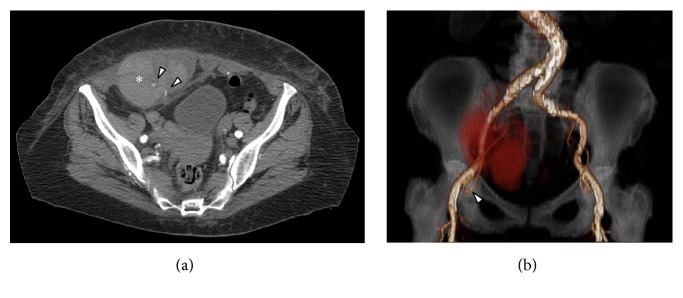
Figure 1.
(a) MD-CT axial view demonstrates the presence of a haematoma into the right rectus sheath (∗) with two spots of active bleeding from distal collaterals of the right inferior epigastric artery (arrowheads). (b) MD-CT coronal Volume Rendering Technique image with right inferior epigastric artery reconstruction (arrowhead) and the haematoma into the right rectus sheath as a red shadow.

