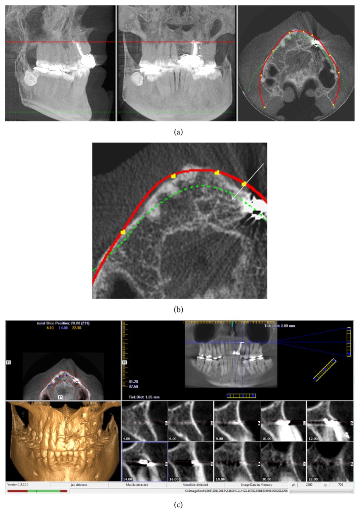
Figure 5.
(a) CBCT (iCAT) imagery taken during original implant assessment by implant clinic. (b) Magnified axial image from (a) showing that a well-demarcated canal is present between the apex of tooth 23 and the nasopalatine canal, white arrow. (c) Multiple foramina are easily seen throughout the apical/nasal region prior to implant placement.

