Fig. 7.
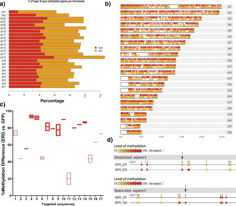
Overexpression of ERG causes global methylation changes. a Percentage of hyper-methylated (red) and hypo-methylated (yellow) regions out of all covered CpGs for each chromosome in the RWPE1-ERG cell line. b Chromosome ideogram showing sites of differential methylation in the RWPE1-ERG cell line. Hyper-methylated CpGs (red) and hypo-methylated CpGs (yellow). c Percentage methylation difference of 17 targeted sequences in the isogenic RWPE1 cells (+/- ERG), measured by the EpiTYPER MassARRAY assay. The thick red outline boxes represent the difference in methylation ratios between RWPE1-ERG and RWPE1-GFP measured on a sequence containing only the targeted CpG. The red boxes with a light outline correspond to an average of methylation ratios on a sequence containing two to four CpGs including the targeted one. For each targeted sequence, the methylation value has been measured in duplicate as shown in (d), except when the composition of the sequence did not allow it (single value for the sequences 2, 8, 10, 12, 13, 16, and 17). d Each CpG is represented by a color-coded circle depending on its methylation ratio. Yellow represents a low level of methylation, red represents a high level of methylation, and grey corresponds to unanalyzed CpGs. The values for the methylation ratio of CpG #4 in sequence 9 determined on both forward and reverse strands are shown in (c)

