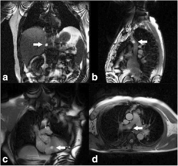
Fig. 3.

CMR fluoroscopy guided right heart catheterization.
a Coronal view with the gadolinium filled balloon at the tip of the catheter in the inferior vena cava (arrow), (b) sagittal view of the superior vena cava, (c) coronal view of the right ventricle, and (d) axial view of the main pulmonary artery bifurcation with the balloon in the right pulmonary artery
