Fig. 2.

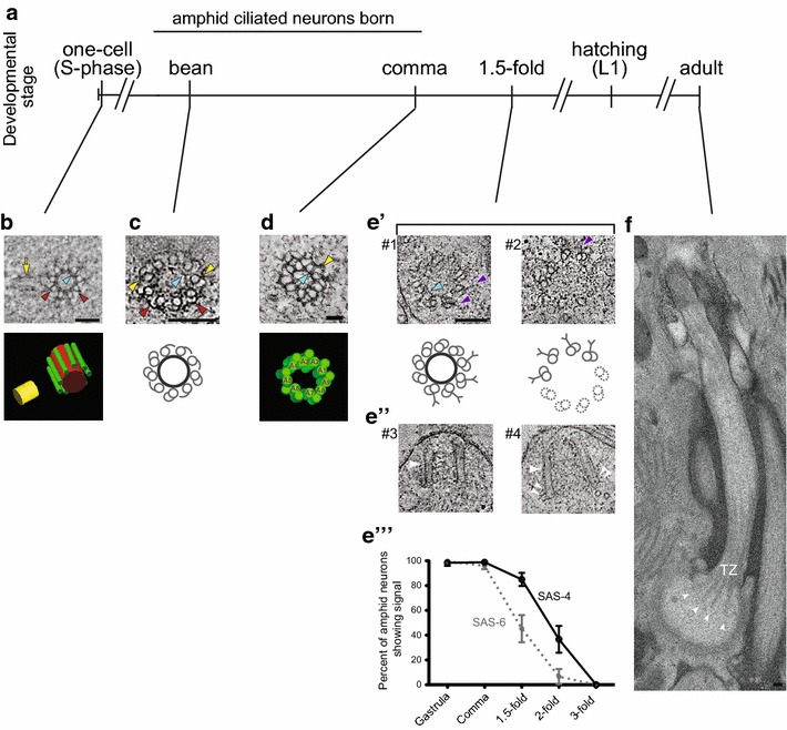
Ultrastructure of C. elegans centrioles and basal bodies. a Developmental timeline of C. elegans and time of birth of amphid sensory neurons. b–e′ Representative cross-section tomographic slices and three-dimensional models or cartoons showing centrioles in the one-cell embryo and basal bodies in amphid neurons at the indicated developmental stages. Note basal bodies containing (#1) or lacking (#2) a central tube in e′. Central tube, sMTs, and the daughter centriole are indicated in red, green and yellow, respectively in the model in b. A1–A9 indicate dMTs in d. Red arrowheads: sMTs with hooks, yellow arrowheads: dMTs, blue arrowheads: central tube, purple arrowheads: Y-links of the transition zone, yellow arrow: daughter centriole. e′′ Longitudinal tomographic slices showing basal bodies/axonemes. Large white arrowheads: basal bodies/axonemes, small white double arrowheads: flared dMTs at the base. e′′′ Quantification of SAS-6 and SAS-4 signals at the amphid sensory neuron basal bodies through embryonic development. f A longitudinal section of the amphid ASE neuron cilium in the adult hermaphrodite. Note flared dMTs at cilia base (arrowheads). TZ transition zone. Images in b are adapted by permission from Macmillan Publishers Ltd: [24]. Images in c, e′ and e′′ are adapted from [25]. Images in d and e′′′ are adapted from [26]. Image in f is adapted from [15]. Scale bars b, c, e′–e′′′, f 100 nm, d 50 nm
