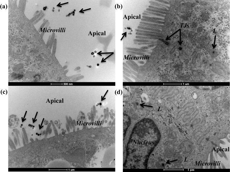
Figure 4.
Transmission electron micrographs of Caco-2 barriers after exposure to 150 nm SiO2-NPs. Caco-2 barriers cultured for 21 days were exposed for 9 h to 100 µg/mL 150 nm SiO2-NPs dispersed in (a,b) the absence of serum, and (c,d) the presence of 10% FBS. The arrows indicate extracellular NPs and some NPs inside vesicles along the endolysosomal pathway. Abbreviations: L, lysosome; TJs, tight junctions.

