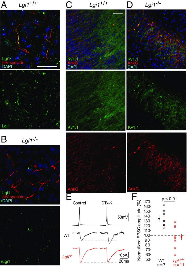
Fig. 6.
Axonal LGI1 regulates excitatory neurotransmission via presynaptic D-type current. (A and B) Representative images from WT and Lgi1−/− brain sections. (A and B) CA3 neurons from WT and Lgi1−/− animals stained with antibodies against the axon initial segment marker βIV-spectrin (red) and LGI1 (green). (Scale bar, 50 µm.) LGI1 staining is detected at axon initial segment in WT mice, whereas no staining is detected in Lgi1−/−. (C and D) CA3 region in WT (C) and Lgi1−/− slices (D) stained with antibodies against the axon initial segment marker ankyrinG (red) and Kv1.1 (green). (Scale bar, 100 µm.) (E and F) Representative synaptic recordings from paired CA3 neurons to evaluate the effects of DTX-K on EPSC amplitude in WT and Lgi1−/− animals (E) and data analysis (F). Error bars represent SEM.

